Our ability to sense change, feel pain, adapt to injuries, and move gracefully depends on a remarkable information highway: the afferent input system. This network of sensory neurons links every tissue in our body to the nerve centers of the spinal cord and brain, forming the crucial communication bridge for healthy reflexes and movement. Whether you are a clinician striving to resolve chronic patient issues, a researcher exploring new frontiers in neurological function, or a patient searching for answers when pain and weakness linger despite treatment, a clear grasp of afferent input unlocks the path to more precise diagnosis, targeted therapy, and lasting relief.
In practical terms, if you step on a nail or touch something hot, your body somehow knows to pull away instantly, even before you're aware. That rapid response is only possible because specialized nerves detect the irritant, convert it into an electrical signal, and rush that message toward your spinal cord and brain. This automatic defense is just one example of afferent input at work - and why understanding its pathway can be the missing piece for those who struggle with chronic pain, muscle inhibition, or stubborn injuries.
This content is for informational purposes only and is not a substitute for individualized medical advice or care. Always consult a qualified healthcare professional for personal diagnosis or treatment.
Introduction to the Afferent Input System
The concept of afferent input sits at the heart of both basic neuroscience and practical approaches like Afferentology. Here’s how it all begins:
Definition and Origins
- The term afferent comes from the Latin afferre, which means "to carry toward." In anatomy and neurology, afferent nerves are those that carry sensory information toward the central nervous system (CNS), specifically the spinal cord and brain.
- These nerves are composed of specialized sensory neurons. Their primary job is to gather raw data - touch, stretch, pressure, pain, temperature, and chemical states - from skin, muscles, organs, and connective tissues.
Distinction from Efferent Output
- Afferent input brings information in, signaling the presence of an outside force or internal change.
- Efferent output sends instructions out from the CNS, controlling muscles or glands to elicit a response - like activating a muscle to contract.
Think of the afferent input system as thousands of couriers, shuttling real-time reports from every corner of your body to your brain’s command center. Without these couriers, movement would be impossible, and the body would have no way to protect itself from harm.
Overview of the Nervous System Relevant to Afferent Input
Understanding the structure of the nervous system is foundational to appreciating how afferent signals travel and where things can sometimes go wrong.
Central vs. Peripheral Nervous System
- Central Nervous System (CNS): Includes the brain and spinal cord. The CNS receives, processes, and interprets sensory data, coordinating the correct output.
- Peripheral Nervous System (PNS): Comprises all nerves that extend from the CNS throughout the body - delivering input to the CNS and carrying output to muscles and organs.
Comparison Table: Afferent vs. Efferent Neurons
| Feature | Afferent (Sensory/Input) | Efferent (Motor/Output) |
|---|---|---|
| Direction | From periphery to CNS | From CNS to periphery |
| Function | Carry sensory signals (touch, pain) | Carry commands (muscle action) |
| Examples | Muscle spindle Ia fibers, cutaneous receptors | Motor neurons to skeletal muscle |
| Tracts | Ascending pathways | Descending pathways |
Sensory Receptors: The System's Frontline
Sensory receptors detect and translate physical or chemical stimuli into nerve signals:
- Mechanoreceptors: Detect touch, stretch, and pressure.
- Nociceptors: Sense pain.
- Thermoreceptors: Respond to temperature changes.
- Proprioceptors: Monitor position and movement in muscles and joints (muscle spindles, Golgi tendon organs).
Functioning of Afferent Nerves and Key Anatomical Pathways
The journey from sensation to perception - and, if needed, to a protective movement - follows sophisticated neural routes. Here’s how the body makes it happen:
Types of Afferent Nerve Fibers
- Ia fibers: Originating in the primary endings of muscle spindles, these are the fastest and most responsive afferent fibers. They are essential for keeping the brain and spinal cord updated about sudden muscle stretch or length changes. When a muscle suddenly stretches (as when you trip), Ia fibers instantly transmit a signal that initiates a reflex contraction, stabilizing posture before conscious thought occurs. A clinical hallmark of their function is their ability to bifurcate (split) at the spinal cord - contributing to both automatic reflexes and messages that ascend toward the brain.
- Group II fibers: Also from muscle spindles, these relay ongoing information about muscle length under steady tension.
- A-delta and C fibers: Conduct pain and temperature sensations; A-delta fibers are faster, handling rapid, sharp pain; C fibers are slower, transmitting dull, aching pain.
- Visceral afferents: Carry information from the organs, supporting vital reflexes and internal regulation.
Ascending Tracts: The Body’s Information Highways
Afferent signals travel along specific ascending tracts:
- Dorsal column-medial lemniscal system: Responsible for fine touch, vibration, and proprioception (body awareness).
- Spinothalamic tract: Handles pain, temperature, itch, and crude touch.
Afferent Pathway Progression (Visualized)
Picture a muscle spindle in your thigh detecting a stretch. The sensory information travels up a specialized nerve, through the dorsal root ganglion (a neural checkpoint), enters the spinal cord, and from there may:
- Trigger a local reflex via direct synapse with motor neurons or interneurons (for immediate protection).
- Ascend toward higher brain centers for conscious recognition and adaptation.
Regulation and Modulation of Afferent Input
The nervous system must balance incoming sensory data. Without this balance, even minor stimuli could overwhelm us; too little input, on the other hand, can cause loss of muscle tone, delayed reactions, or undetected injury.
Presynaptic Inhibition and Clinical Importance
Presynaptic inhibition is the nervous system’s way of adjusting the volume on incoming signals. Certain interneurons (notably GABAergic neurons) release neurotransmitters that dampen the transmission of signals along afferent fibers before those signals reach the next neuron. The outcome keeps responses proportional and prevents reflexes from firing unnecessarily.
Through this process, sensory input that is irrelevant or non-dangerous is effectively "turned down," so we don't experience constant, distracting reflexes. For example, when your shirt brushes your skin, you aren't continually jumping or moving away.
Central and Peripheral Modulation
- Central modulation: The brain and spinal cord select and prioritize which signals to act on. Endorphins, for example, can suppress pain signals during emergencies.
- Peripheral changes: Inflammation or tissue damage lowers the threshold required for afferent neurons to fire, which can make ordinary touch feel painful (allodynia).
Effects of Spinal Cord Injury and Maladaptive Rewiring
After injury, nerve fibers can sprout and create new, sometimes chaotic connections. This can lead to hyperreflexia (exaggerated reflexes) or persistent, uncontrolled muscle contraction and pain. Synaptic changes can also cause prolonged excitatory postsynaptic potentials (EPSPs), where a single input generates drawn-out or repetitive nerve firing.
Interneurons: Integration at the Crossroads
Interneurons are small but powerful nerve cells in the spinal cord and brain. They act as relays, connecting afferent (input) neurons with efferent (output) neurons. This network makes possible fast reflexes, intricate movement coordination, and adaptive learning from repeated exposure or injury.
Clinical Significance of the Afferent Input System
The role of afferent input extends from everyday safety to challenging clinical scenarios where persistent pain, weak muscles, or unexplained injury risk seem immune to treatment.
The Withdrawal Reflex: A Blueprint for Survival
When a sharp object touches your skin, nociceptors (pain receptors) fire an afferent signal to the spinal cord. This information is relayed instantly to motor neurons and interneurons, activating the Withdrawal Reflex. The affected muscles contract to pull you away from danger - no conscious intervention required at first.
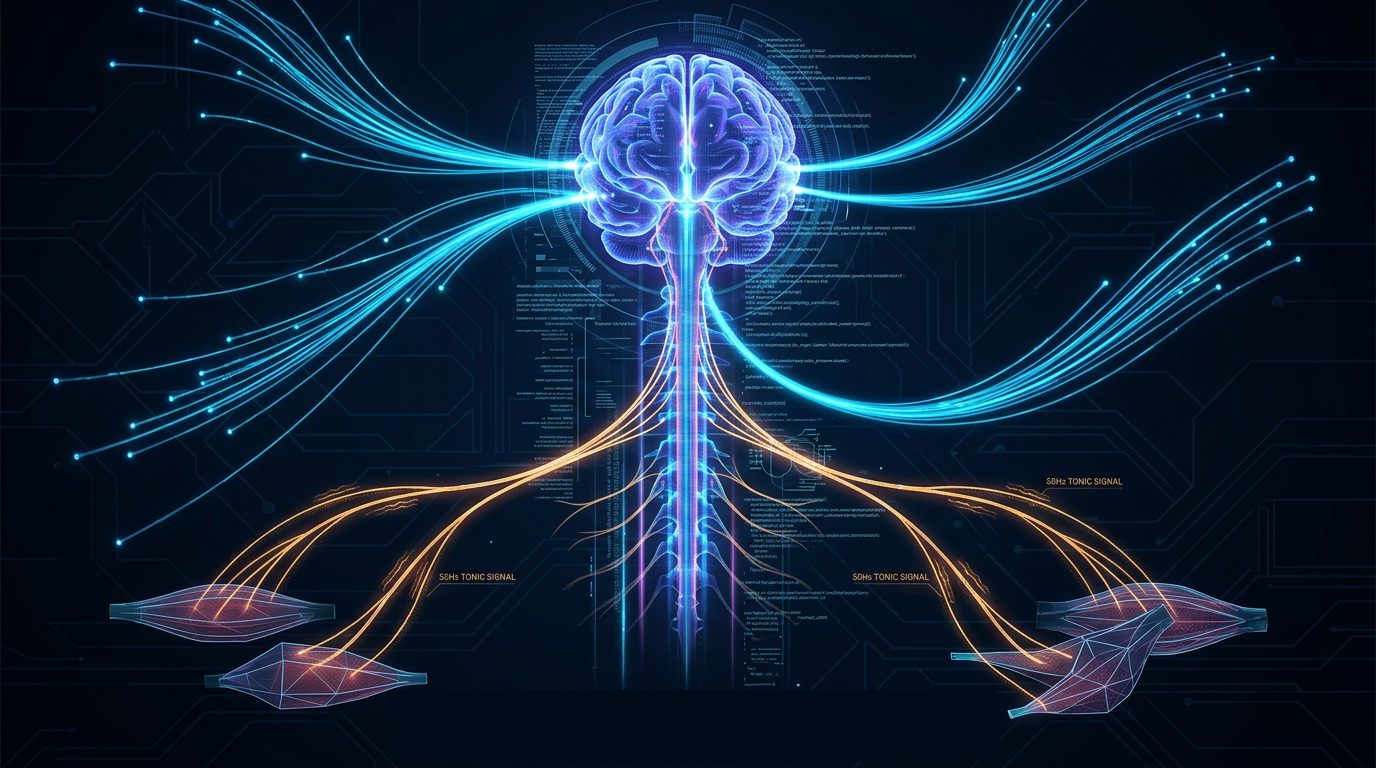
Muscle Tone and the 50Hz Resting Benchmark
Muscle tone is the default tension held in our muscles - not enough to cause movement, but always primed for action. Optimal tone depends on continuous, balanced afferent input, with muscle spindles sending regular pulses at specific frequencies (notably around 50Hz). Disruption (from injury, scarring, or nerve interference) can lead to muscle inhibition, poor readiness, and a cascade of compensation elsewhere in the body.
Hidden Irritants and the Origins of Nerve Interference
Often, patients experience muscle weakness or chronic pain that defies traditional explanations. Afferentology highlights a range of often-overlooked sources of nerve interference - sometimes described as "software corruption" - that can perpetuate pain and inhibit muscle function:
- Active scar tissue: Healed wounds may hide nerves firing incorrectly, sending a constant low-level pain signal even when the tissue appears fine.
- Dental interference: Jaw tension, old fillings, imbalanced bite, and related dental issues can signal the entire body, not just local structures.
- Joint dysfunction: Restricted motion irritates articular receptors, which then trigger muscle inhibition - think of it as a sticky joint "locking out" surrounding muscles.
- Visceral referral: Internal organ issues (such as liver, bowel, or reproductive organs) often refer pain to distant body regions, altering movement and muscle readiness.
- Fascial adhesions: Stiffened, thickened areas of connective tissue block normal signal transmission and feedback.
Clinical Tools: Precision Muscle Testing and Beyond
Precision Muscle Testing is one of the central diagnostic tools in Afferentology. By targeting specific muscle groups and applying clinical protocols, practitioners can map which areas of the nervous system are operating as expected and which suffer from inhibition or interference.
Testing allows for real-time feedback. Once the principal irritant (scar, joint, dental, or visceral issue) is addressed, muscular function may return almost immediately - a phenomenon witnessed repeatedly in documented case studies.
Bridging Research and Practice
A growing body of evidence supports the need for a thorough afferent system assessment in patients with persistent muscle inhibition or unexplained pain. Simon King and the Association of Certified Afferentologists emphasize protocols rooted firmly in neuroscience, offering fresh solutions for clinicians and hope for those whose symptoms have resisted conventional care.