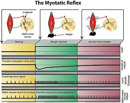
For years I taught that a resting muscle hums along at a steady 50Hz. I was wrong, and a textbook diagram was partly to blame.
You'll know the one. Muscle, spindle, spinal cord, and a neat row of tick marks showing the alpha motor neuron firing away at rest. Add a weight, the ticks speed up, the muscle contracts, and everything settles back to normal.
It's a lovely picture. It just isn't what a relaxed muscle does.
What a Relaxed Muscle Is Really Doing
In 1986, Ribot and colleagues managed something extraordinary. They recorded single motor fibres in awake human volunteers. When the muscle was relaxed, the alpha motor fibres, the ones that make muscle contract, were completely silent.
So the stiffness you feel in a resting muscle isn't the nervous system holding it taut. It's mostly passive: connective tissue, the elastic protein titin, and a few cross-bridges that stay attached while the muscle is still.
But the nervous system wasn't idle. The same study found the gamma (fusimotor) fibres firing at rest. Gamma motor neurons don't move the muscle. They supply the tiny muscle fibres inside the muscle spindle, the sensor that reports stretch.
They responded to a clap of hands, to mental arithmetic, even to the subject listening to instructions. One subject could switch a unit off at will.
A relaxed muscle is mechanically quiet but neurologically awake.
The Muscle Spindle: The Segment's Main Reporter
Many of us were taught that joint receptors tell the brain where a joint is. The recordings say otherwise.
When Burgess and Clark recorded nerve fibres from the cat knee joint in 1969, fewer than 5% fired in the middle of the range. Most only woke up near the ends. Some of the mid-range "joint" signals turned out to come from muscle spindles whose nerves happened to travel in the joint nerve.
Spindles fire throughout the range and keep firing during contraction. So most of what the nervous system hears from a moving spinal segment, it hears from the muscles, not the joint.
Afferent input is the battery of your brain. Around the spine, most of that charge comes from muscle.
Gamma: The Tensioner Inside the Sensor
When a muscle shortens, its spindles should go slack and fall silent. Gamma prevents that by contracting the ends of the spindle's own fibres, keeping the sensor loaded.
It's a tug-of-war. A 2020 model by Blum and colleagues showed that if alpha shortens the muscle faster than gamma can tension the spindle, the spindle goes quiet. If gamma wins, it fires more. Somewhere in between, nothing changes.
That explains an old puzzle from Edin and Vallbo. Two spindles in the same human finger muscle, during the same contraction, producing the same force. One fired more. The other fired less.
A Sensor with a Memory
Spindles also remember. After a few seconds of stillness, cross-bridges inside the spindle settle into place, so the first stretch meets stiff resistance and fires a large burst. Stretch again straight away and the burst is much smaller. The memory fades over roughly 5 to 10 seconds.
How many of us control what the patient did in the ten seconds before we test them?
Active and Passive Are Different Questions
When you stretch a relaxed muscle, alpha is silent, the spindles report the stretch, and what resists you is mostly tissue. A quick stretch adds some reflex resistance. A slow one adds very little.
When the patient moves, alpha is firing, gamma keeps the spindles reporting, and the opposing muscle is told to let go.
Motion palpation asks the passive question. Your patient lives with the answer to the active one.
Three Words Worth Getting Right
Before the next article, a note on language. Many of us use "facilitated" to mean a muscle that tests strong. In neurophysiology it means something else: a motor neuron pushed closer to firing, whether or not it actually fires.
So I'll use three plain words. Normal: a muscle that switches on and lets go appropriately. Inhibited: less able to switch on than it should be. Overactive: switches on too readily or won't let go, usually because it has lost the inhibition it would normally receive.
Next time: what happens to a spinal segment when one of its muscles stops working.
To your strength!