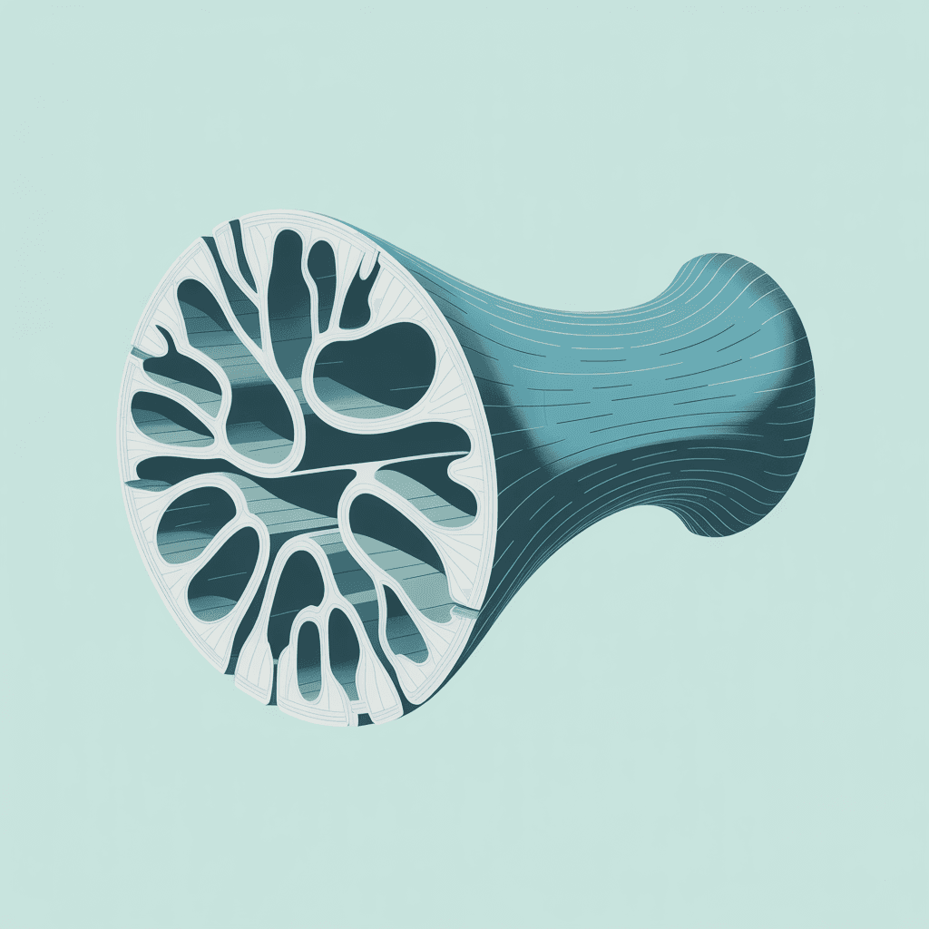
Osteoporosis management is dominated by two interventions: pharmaceutical agents that alter bone remodelling kinetics, and weight-bearing exercise to apply mechanical loading to the skeleton. Both approaches operate on the correct principle — that bone density responds to mechanical demand — but the second approach is frequently undermined by a factor that is never assessed. Exercise generates bone loading signals through muscle contraction. The compressive and tensile forces applied to bone through muscular activity are what drive osteoblastic activity and maintain cortical density. When muscles are chronically inhibited by afferent withdrawal, the loading signal to bone is chronically reduced, and no amount of exercise prescription can compensate if the motor neuron supply to the relevant muscles is compromised by a withdrawal reflex.
Afferentology frames osteoporosis not as a calcium metabolism problem or a hormonal deficiency alone, but as the eventual structural consequence of a muscular loading failure. A patient who has been prescribed weight-bearing exercise for bone density and whose bone density has continued to decline is a patient whose musculoskeletal system may not be generating adequate loading signals to the skeleton — not because they are not exercising but because the motor neuron supply to the relevant muscles has been reduced by an afferent withdrawal reflex that was never identified.
Bone Remodelling and the Muscular Loading Signal
Bone remodelling is regulated by mechanical loading via the piezoelectric properties of bone and the mechanosensitivity of osteocytes. Osteocytes — the most abundant bone cells — detect mechanical strain and regulate the balance between osteoblastic bone formation and osteoclastic bone resorption. When mechanical strain is adequate and appropriately varied, osteoblasts dominate and bone density is maintained. When strain is reduced — as occurs in immobilisation, microgravity, or chronic muscular inhibition — osteoclastic activity dominates and resorption exceeds formation. Bone density decreases. The threshold for adequate mechanical strain is determined by the force generated through muscular contraction, not simply by the act of weight-bearing.
The Biomechanical Reality of Muscle Pull: Biomechanical tracing studies show that internal muscle contractions represent the vast majority of adaptive bone responses. For instance, over 70% of the mechanical bending forces generated inside the femur during walking are developed purely by the internal action of pulling muscles, while less than 30% come from the gravitational impact of body weight hitting the ground. Furthermore, this dynamic process is driven by mechanotransduction, where structural micro-deformations open specialized, mechanosensitive Piezo1 ion channels on bone cells, flooding them with calcium to activate bone-building osteoblasts.
A patient whose gluteus medius is chronically inhibited by a lumbar disc afferent source does not generate adequate compressive strain through the femoral neck during walking. Their weight-bearing exercise produces insufficient osteocyte and Piezo1 stimulation to maintain femoral neck density. The hip fracture that eventually occurs is attributed to osteoporosis — to a bone density that was below fracture threshold. What is not attributed is the decade of gluteus medius inhibition that progressively reduced the loading signal to the femoral neck, allowing the density to fall below that threshold while the patient was apparently "active." The muscular software failure is the upstream event. The bone density loss is the downstream consequence.
"Bone density is not maintained by weight-bearing. It is maintained by muscular force generation. A patient who walks every day but whose hip abductors are chronically inhibited is not adequately loading their femoral neck with every step. The bone responds to what the muscles actually do, not what the exercise prescription says they should do."
What Is Driving the Muscular Inhibition That Reduces Bone Loading
The human motor neuron continuously fires at an involuntary baseline frequency of approximately 50Hz to maintain healthy resting muscle tone. This persistent 50Hz "hum" acts as a continuous low-intensity, high-frequency stimulus perfectly tuned to optimize osteoblast proliferation and maintain structural baseline density. The afferent sources most commonly producing the chronic muscular inhibition that drops this frequency below the 50Hz benchmark include:
- Lumbar disc afferents reducing lower limb muscle output: The gluteus medius, quadriceps, tibialis anterior, and calf muscles are all supplied from the lumbar cord segments. Chronic, low-level disc afferent activity at L3-S1 — which may produce minimal or no pain — reduces the 50Hz resting tone of these muscles and the force they generate during loading activities. The femur, tibia, and calcaneum are consequently under-loaded with every step, and bone remodelling shifts toward resorption over years and decades.
- Abdominal scar afferents inhibiting paraspinal muscles: The thoracic and lumbar paraspinal muscles generate the axial compression forces that load the vertebral bodies. When these muscles are chronically inhibited by abdominal scar afferents — from previous abdominal or spinal surgery — the vertebral loading during movement is reduced, and vertebral body density decreases. The vertebral fractures that characterise advanced osteoporosis are occurring in bone that was chronically under-loaded by inhibited paraspinal muscles.
- Upper limb surgical scars reducing arm and shoulder muscle tone: Bone density in the proximal humerus, radius, and ulna depends on the forces generated by the shoulder girdle, rotator cuff, and forearm musculature. Chronic inhibition of these muscles — from cervical disc, thoracic outlet, or shoulder and chest wall scar afferents — reduces the loading signal to the upper limb bones. This local coupling is why grip strength directly correlates with upper limb bone density, completely independent of systemic hormone status.
- Post-menopausal hormonal changes increasing afferent sensitivity: Oestrogen modulates the mechanoreceptor sensitivity of joint and ligament afferents. Post-menopausal oestrogen reduction increases afferent sensitivity, making existing withdrawal reflex sources more clinically potent and increasing the magnitude of the muscular inhibition they produce. A withdrawal reflex source that was subclinical in a pre-menopausal patient may become clinically significant post-menopausally — producing the combination of accelerated bone density loss and muscular weakness that characterises post-menopausal osteoporosis.
The Ultimate Proof of Neurological Control: To understand the dominance of the nervous system over human architecture, contrast post-menopausal changes with spinal cord injuries (SCI). While ambulatory post-menopausal women lose bone mineral density (BMD) at an average rate of roughly 2% per year, paraplegic patients experience a catastrophic shutdown of their motor software, losing approximately 1% of their lower limb BMD per week. Within six months of complete neurological disconnection, a paraplegic can lose up to 40% of their lower limb bone mass, leading to severe microarchitectural devastation (up to 62% greater structural empty spacing in trabecular bone compared to estrogen-free women). This proves that local osteoporosis is fundamentally driven by a rapid failure of neurological and muscular software.
The Software Test: Protective Reflex Testing
PRT in osteoporosis management is applied as an audit of the motor neuron supply to the muscles responsible for loading the most vulnerable skeletal sites. For the hip, this means testing the gluteus medius, gluteus maximus, and quadriceps — the muscles whose loading of the femoral neck and proximal femur determines hip fracture risk. For the spine, this means testing the paraspinal muscles at the thoracic and lumbar levels. For the wrist, this means testing the shoulder and forearm musculature. If any of these muscles are inhibited, the withdrawal reflex source is identified and addressed before the exercise programme is prescribed. The exercise programme that follows this neurological restoration is working with a musculoskeletal system capable of generating the loading forces that bone remodelling requires.
In practice, patients with established osteoporosis frequently show multiple muscular inhibition patterns that have been present for decades. Source identification and resolution does not reverse existing bone density loss — the structural damage is real. What it does is stop the progression: restoring the loading signal to bone through muscular tone restoration removes the mechanical driver of continued bone resorption. Combined with appropriate pharmaceutical management where indicated, this neurological restoration represents a genuinely comprehensive approach to osteoporosis management that no current pharmacological protocol addresses.
Clinical Takeaways
- Weight-bearing exercise is effective only if the muscles are generating adequate loading force: A patient who is prescribed walking or resistance exercise for bone density should have their relevant musculature assessed with PRT before the prescription is made. Exercise into chronic muscular inhibition generates reduced loading signals and suboptimal bone remodelling responses.
- The paraspinal muscles are the primary loading structure for the vertebrae: Vertebral osteoporosis and vertebral compression fractures occur in bone that is chronically under-loaded. Paraspinal muscular inhibition from abdominal or spinal scars is a frequently overlooked contributor to vertebral bone density loss that standard osteoporosis management does not assess.
- Post-menopausal osteoporosis has a neurological amplification component: The reduction in oestrogen post-menopausally increases the clinical potency of existing withdrawal reflex sources. PRT assessment in post-menopausal patients with accelerating bone density loss should identify and address these sources as part of a comprehensive management approach.
- Fracture risk is not only a bone density variable: A patient with a withdrawal reflex pattern that chronically inhibits the hip abductors and quadriceps has increased fall risk — because those muscles protect against uncontrolled movement — and reduced bone density at the hip — because those same muscles are not loading it adequately. The neurological audit addresses both components of fracture risk simultaneously.
Osteoporosis is not simply a bone problem. It is a loading problem. And the loading problem has a neurological explanation.
The muscles are not generating adequate force. The bone is not receiving adequate mechanical signal. The bone resorbs. Address the muscular inhibition that is reducing the loading signal, and the bone has a reason to maintain its density. Exercise is essential. But exercise into inhibition is not the same as exercise with full motor neuron supply.