In the early 1960s, a Swedish neurophysiologist pushed a fine tungsten needle through his own skin and into his own ulnar nerve. He wanted to hear what a living human nerve sounds like.
Before we get to what he heard, let's look at what he was listening to.
Most of the Afferent Traffic Is Coming In
We tend to think of nerves as the brain's way of sending orders out to the body. The numbers say otherwise.
When Gesslbauer and colleagues counted the axons in the nerves of the human arm, they found around 350,000 axons leaving the spinal cord for the upper limb, and only 10% of them were motor. In every nerve they examined, sensory axons outnumbered motor axons by at least nine to one.
Even a nerve going to a single muscle is mostly listening. The cat soleus nerve has roughly 450 myelinated axons, and only about a third of them are alpha motor neurons carrying commands to the muscle. Another 115 are gamma motor neurons supplying the muscle spindles. The rest bring information back.
Afferent input is the battery of your brain. The wiring proves it.
The Anatomy of a Spinal Nerve
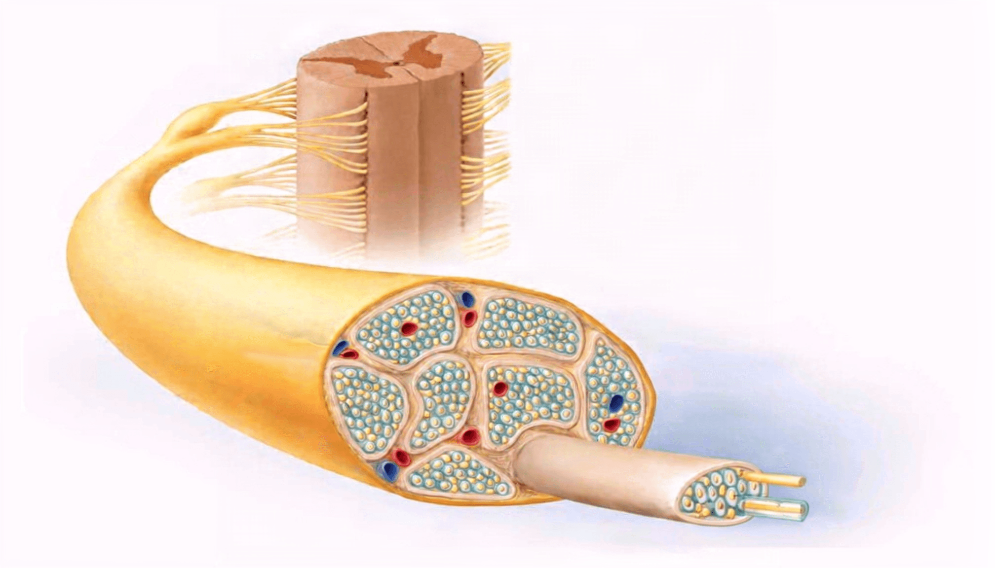
Each spinal segment sends out a pair of spinal nerves, one on each side. Each nerve starts as two separate roots.
The dorsal root, at the back, is sensory. It carries information coming in from skin, muscles and joints. The cell bodies of these sensory neurons sit in a small swelling just outside the cord called the dorsal root ganglion.
The ventral root, at the front, is motor. It carries alpha motor neurons to the muscle fibres and gamma motor neurons to the spindles.
Two hundred years ago, Bell and Magendie worked out this split: in at the back, out at the front.
The two roots join in the intervertebral foramen to form the mixed spinal nerve. It's barely a centimetre long. Almost immediately it divides again:
- the dorsal ramus runs backwards to supply the paraspinal muscles, the facet joints and the skin of the back;
- the ventral ramus runs forwards to supply the limbs and the front and sides of the trunk;
- a tiny meningeal branch turns back into the spinal canal.
Inside, the nerve is packaged like the cable behind your television. Each axon sits in its own sleeve of connective tissue, the endoneurium. Axons are bundled into fascicles wrapped in perineurium, and the whole nerve is wrapped in a tough outer sheath, the epineurium.
Why Nerve Signals Don't Jump Between Fibres
Thousands of axons are packed together, many carrying impulses at the same moment. So why doesn't a signal heading out to a muscle leak into the fibre beside it?
Because each axon makes its own signal. An action potential isn't a current pushed down a wire. It's regenerated at each point along the axon by ion channels opening in its own membrane.
The tiny current that leaks outside an active axon spreads out into the surrounding fluid and dissipates. By the time it reaches a neighbouring axon, it's far too weak to push that axon's membrane to threshold. Myelin and the connective tissue sleeves add more insulation.
Direction is set at the junctions, not in the cable. An axon stimulated in the middle will conduct both ways. But synapses only pass signals one way, so sensory traffic always ends up flowing in and motor traffic out.
There's one important exception. When myelin is damaged, bare axons can lie against each other, and one can excite its neighbour directly. This is called ephaptic cross-talk.
In trigeminal neuralgia, where an artery usually compresses the nerve root, the compressed area shows patches of lost myelin with bare axons lying directly against each other. Separating the vessel from the nerve brings rapid relief in most patients, probably because it pulls those axons apart and stops the excitation spreading between them.
Injured nerves can do the same. In rats, several weeks after a sciatic nerve injury, stable electrical cross-talk formed between pairs of damaged sensory and motor axons.
Crossed wires are possible, but they're a sign of damage. A healthy nerve keeps its traffic separate.
How We First Measured a Nerve Impulse
For most of history, nobody knew what travelled along a nerve.
In the mid-1800s, du Bois-Reymond showed the nerve impulse is an electrical wave travelling along the nerve, and Helmholtz made the first measurements of its speed. Helmholtz's figure of about 27 metres per second in frog nerve was far slower than anyone expected. His own father was reportedly sceptical.
By the 1920s, Edgar Adrian was using radio amplifiers to record single nerve fibres, and he found that sense organs fire not single impulses but rapid trains of them. Each impulse in a fibre is the same size. A stronger stimulus produces impulses more often, not bigger ones.
Erlanger and Gasser needed something faster to record these brief signals. They built their own cathode ray tube from an Erlenmeyer flask coated in fluorescent paint, creating the first practical oscilloscope for recording nerve signals. With it, they showed that nerve fibres don't all conduct at the same speed: larger fibres conduct faster than smaller ones. The work identified three main types of nerve fibre and won them the 1944 Nobel Prize. Those A, B and C fibres are still how we classify nerves today.
They also showed that many properties of a fibre depend on its conduction speed, including how sensitive it is to pressure. The large, fast fibres carrying touch, muscle spindle signals and motor commands are the first to fail under compression. The smallest fibres are the most resistant.
All of this, though, was done on animal nerves.
Microneurography: Listening to a Living Human Nerve
Before the late 1960s, impulses in peripheral nerves had only been recorded in animals, by dissecting the nerve and splitting it into strands. Putting a needle into a human nerve was widely thought likely to cause permanent damage.
Which brings us back to Sweden. Karl-Erik Hagbarth and Åke Vallbo, working at the Academic Hospital in Uppsala, repeatedly needled each other's nerves to find the best electrode and insulation, and settled on tungsten coated in epoxy. Hagbarth was the first to try it, on his own ulnar nerve. For about two years they experimented on their own nerves, checking carefully for damage.
Then, in a burst of papers, they documented muscle afferents, muscle efferents and skin afferents in awake humans.
This is how it's done today. The electrode has a shaft about 100 to 200 micrometres across and a tip of 1 to 5 micrometres, insulated to the tip. It goes a few millimetres through the skin, and weak electrical pulses through it help locate the nerve. The investigator then advances it with small, slow movements while listening to the nerve's activity through an amplifier.
When a single fibre is isolated, its receptor is found by gently probing the skin or muscle until it fires. Many laboratories now use high-frequency ultrasound to watch the electrode entering the nerve.
The great strength of the method is that the subject is fully awake and can take part in tasks that need attention while their nerve fibres are recorded. The same electrode can also stimulate a single fibre so the person can report what they feel. That approach has recently been revived and combined with high-resolution brain imaging.
Almost everything we know about human muscle spindles comes from this one technique.
What Microneurography Can't Do
It has real limits, and they matter.
It only reaches nerves you can get a needle into. Recordings are made from accessible peripheral or cranial nerves, such as the median, ulnar and peroneal. Nobody records from a spinal nerve inside the foramen, or from the roots. Everything we say about traffic in the spinal nerve itself is inferred from limb nerves and animal work.
It sees a few fibres at a time. A good session might yield a handful of units from a nerve containing tens of thousands. That's a very small sample.
It favours the big fibres. Hallin found that standard tungsten electrodes preferentially recorded the myelinated fibres in a fascicle. Small fibres need special electrodes and patience.
It hates movement. The electrode tip sits within a few micrometres of an axon. Large or fast movements dislodge it, so most recordings use small, slow or isometric movements, not the activities we care about clinically.
It's slow and highly skilled. Only a handful of laboratories worldwide do single-unit microneurography, because of the ethical, practical and mental challenges, and it takes extensive training to master.
Identifying a fibre is often indirect. You find what a fibre does by what makes it fire. Gamma motor fibres are especially hard to pin down, which is why direct human recordings of them are still counted in single figures.
What This Means for Afferent Input
A spinal nerve is mostly a listening device. Nine in ten fibres are carrying information in. The dorsal ramus serves the very muscles and joints we assess every day.
Each fibre keeps its message to itself unless it's damaged. And under pressure, it's the large fibres serving touch, muscle sense and movement that fail first.
Microneurography is one of the great achievements of human physiology, and it began with two men prepared to needle their own nerves. But it hears only a few fibres, in nerves far from the spine, in a body that has to keep still.
When anyone tells you exactly what's happening inside a patient's spinal nerve, it's worth asking how they know.
To your strength!