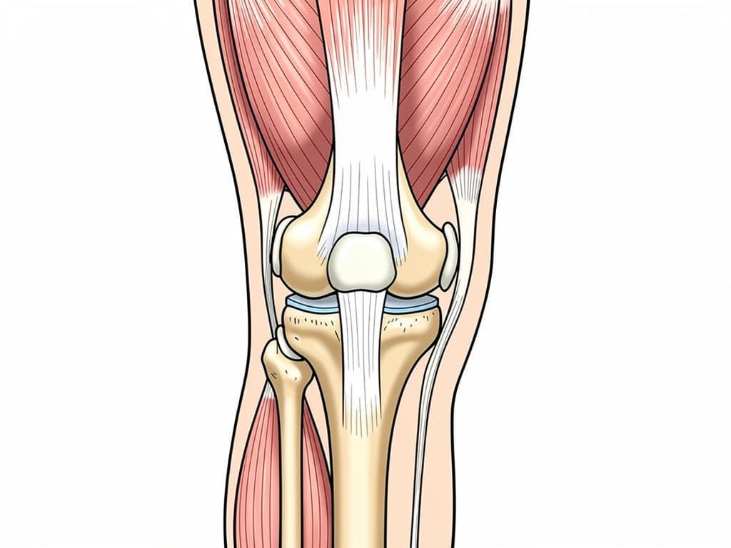
The quadriceps are the most important soft-tissue structures in the knee. Not the cruciate ligaments, not the menisci, not the patellar tendon — the quadriceps. Their eccentric contraction controls knee flexion under load, decelerates descent during stair-climbing, and absorbs ground reaction force during every step. When these muscles are neurologically inhibited, force transfers directly to joint surfaces, cartilage, and ligaments. The joint degrades. The standard clinical response, prescribing quadriceps strengthening, is a reasonable idea that fails consistently because the reason the quadriceps are inhibited is never addressed.
Afferentology frames this differently. A patient who cannot voluntarily activate their quadriceps adequately despite months of physiotherapy is not presenting with a muscle problem they are presenting with a control problem. Something upstream is reducing the motor neuron output to the quadriceps group via the femoral nerve and the L2, L3, and L4 cord segments. Until that source is identified and removed, any rehabilitation programme is destined to failure or sub-optimal results.
Quadriceps Inhibition: The Myotatic Reflex and Its Dysfunction
The quadriceps maintain their resting tone through the myotatic reflex, which responds to stretch and tension with an automatic contraction that protects the knee joint from uncontrolled load. During loading activities lke walking, running, descending stairs, the quadriceps eccentrically resist knee flexion. This is not a conscious action; it is a reflex which depends entirely on the integrity of the motor neuron supply from the L2-L4 cord segments. When the motor neurons supplying the quadriceps are inhibited by increased pre-synaptic inputs, the quadriceps cannot perform their protective role. The knee moves badly, into flexion under load faster than the muscles can respond so the force is transmitted directly to the articular cartilage, the menisci, and supporting ligaments. The joint is being destroyed by a neurological failure, and the mechanism is invisible to standard assessment techniques.
The clinical picture is distinctive once you know what to look for: a patient who describes knee pain on stairs or during prolonged walking, who has been compliant with their physiotherapy programme but who still cannot generate adequate force on knee extension testing. The muscle is physically present but the nerve supply is compromised. Standard testing, even isokinetic dynamometry, measures the output of the compromised motor neuron supply and concludes the muscle is weak. Protective Reflex Testing is a type of manual muscle testing which tries to identify a weakness in the motor neuron supply that needs explaining.
"The quadriceps may be weak due to a muscle or a nerve issue. These are not the same problem, and they do not have the same solution. One responds to exercise, the other responds to finding the abnormal afferent source."
What Is Driving the Inhibition?
The afferent inputs that most commonly inhibit the quadriceps group are varied and frequently remote from the knee:
- Lumbar disc afferents at L3-L4: The femoral nerve derives primarily from L2, L3, and L4. A disc herniation or facet irritation at L3-L4 may generate muscle changes which reduces motor neuron output to the entire quadriceps group. The patient may have minimal or no back pain, but the knee is unprotected by a problem in the lumbar spine. The lumbar disc is the source; the knee is the consequence.
- Anterior hip capsule and inguinal afferents: The anterior hip capsule and the inguinal region are innervated by branches of the femoral and lateral femoral cutaneous nerves. Stress or pathology from an anterior hip replacement, an inguinal scar, or a tight iliopsoas generates afferent input that the spinal cord interprets through the same cord segments that supply the quadriceps, producing partial inhibition.
- Knee joint afferents following injury or surgery: Joint afferents from the knee itself, when carrying nociceptive signals from a sprain, a meniscal tear, or post-surgical healing tissue generate an ipsilateral withdrawal reflex that inhibits the quadriceps. This is the neurological basis of arthrogenic muscle inhibition. The joint signals threat; the cord reduces motor neuron output to the muscles that load that joint. This is entirely logical — and entirely counterproductive when the joint has healed but the afferent signal persists.
The Software Test: Protective Reflex Testing
PRT of the quadriceps group involves testing the muscle in different positions to make sure that it works responds adequately to an external force all of the time. Many times weakness of the knee only shows up when the knee is slightly unlocked, so we test it in that position. Weakness of the knee in this position is common in osteoarthritis of the knee and often presents as a patient who struggles to walk up and down stairs.
The clinical contrast is immediate and objective. With quadriceps inhibition, the patient cannot generate adequate resistance against the practitioner's force. After removing irritations the same muscles resist with full force and can easily adapt to resist a variable force. No exercise needed. Only the afferent input to the spinal cord changed.
Quadriceps failure is usually not a muscle problem, it is a functional motor neuron problem, usually with an identifiable source.
The knee is being destroyed because the control mechanisms that protect it have been compromised. Find the inhibition, restore the motor neuron supply and the quadriceps can do their job. Until then, every exercise programme is treating the wrong problem.